How to cure rickets
Rickets is a disease that develops due to lack of vitamin D also known as hypovitaminosis i.e. phosphorus disorders and calcium metabolism, leading to functional disturbance of the Central Nervous System (CNS) as well as changes in bones i.e. joint and muscle systems, accompanied by the development of polyvalent hypovitaminosis.
Inadequate intake of calcium from food or violation of its absorption in the intestine is accompanied by a decrease in calcium levels, and its ionized fraction in serum. Hypocalcemia stimulates production and secretion of parathyroid hormone which leads to reduction of calcium from bones and increase its level in serum as well as increasing its reflection in the kidneys and increase the synthesis of calcium in the kidneys which is known as Triolet. Homeostasis of phosphate is often regulated by the kidneys i.e. they are absorbed in the intestine almost completely, and their excretion in the urine determines their level in the blood. Excessive absorption of phosphate in the intestine leads to a reduction in serum ionized calcium and increased parathyroid hormone secretion, manifested phosphaturia accompanied by a decrease in phosphate content in the serum and the increase in the amount of calcium. Hypophosphataemia blocks the secretion of parathyroid hormone and activates the synthesis of the calcitriol in the kidneys. This connection enhances the absorption of phosphate in the intestine. Understanding the metabolism of vitamin D is necessary for the understanding of rickets.
The skin contains an enzyme that is under the influence of ultraviolet radiation is converted into vitamin D3. Unavailability of skin to ultraviolet radiation due to smog or clothing also leads to rickets.
There are seven natural substances containing vitamin D with activity but in medical practice they use only 2 of them known as vitamin D2 – called “calciferol” and D3 which is called “cholecalciferol”. In the organism the vitamin itself does not function but its active metabolites, in particular calcitriol.
Calcitriol and other vitamin D metabolites penetrate cell membranes and in cells of organs targeting and interact with specific receptors, forming a complex with them, penetrating into the nucleus of cells and initiates protein synthesis in them. Proteins may be specific (proteins, calcium binding – BSK) and nonspecific – collagen, alkaline fosfatoza. Formed in the brush rim mucosal cells of intestine alkaline fosfatoza participates in an active capturing of calcium ions from the intestinal lumen into the cell with which it interacts with the protein binds calcium, facilitating the passage of calcium through the gut wall into the blood.
Under the influence of metabolites of vitamin D in the intestinal-specific form factors which are also necessary stuff for the absorption of phosphate and magnesium ions. The protein binds calcium circulating in the blood, helps transport calcium in the tissue. In the bones under the influence of calcium Triola is synthesized protein binding calcium, alkaline fosfatoz and normal collagen structure. In the diaphysis kaltsiytriol intensifying resorption of bone tissue it is demonstrated in increasing intake of calcium acetate in blood plasma, which in the kidney is filtered into the primary urine, and from it and then reabsorbed (with normal vitamin D in the body), maintaining a healthy level and citrate, calcium and blood . Resorption of bone with normal vitamin A in the body occurs with a low rate in terms of the resumption of bone tissue. In the kidneys under the influence of calcitriol and is synthesized protein binding calcium, alkaline phosphatase, required for the reabsorption in the proximal tubules of kidneys of calcium, sodium, phosphate, amino acids.
Deficiency of mineral salts violates bone mineralization. Their failure in the areas of growth, shown slowdown and lag bone age is called rickets. The lack of mineralization of trabecular bone means leading to a high content nonmineralized osteoid which is called osteomalacia. Rickets is sick only a growing child and the process in this case is localized mainly in the epiphyses and osteomalacia can occur at any age. All patients revealed osteomalacia rickets, but not all patients with osteomalacia rickets develops.
The disease can occur as a background of deficiency of calcium or phosphorus. Since the ions of calcium and phosphorus are the mineral part of bone, the lack of any of them in the extracellular fluid bathing the mineralized surface, leads to rickets.
Periods of disease are: initial, peak, recovery, residual effects.
The initial stage of the disease accounts for up to the third month of life and shows changes in the nervous system: the child becomes irritable, often crying, sweating, there is a disorder of the fecalium and balding. This period lasts 2 – 3 weeks. In the blood decreases the amount of phosphorus with alkaline phosphatase is activated. In the midst of illness the changes in bone occur: strain softening of skull bones, soften the edges, sternum forward. As a result we see the chest being deformed, deformed limb bones, bone tissue becomes brittle and along with rickets osteoporosis. The X-ray shows not just proper bones but the contours of the bones lubricated. Tubular bone-shaped, fringed edge of the metaphysis. When rickets II – III degree enlarged liver and spleen, expressed anemia.
In acute course has been a rapid increase in symptoms of rickets. Acute course in children during the first months of life, especially in preterm good gain in weight.
Pathogenetic mechanism lies in the violation of phosphate reabsorption in the proximal tubules of the kidneys, the transformation of the inactive metabolite of vitamin D in the active form. Left untreated, the overall body length of an adult does not exceed 130 – 160 cm.
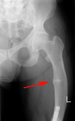
Radiographic signs are to enhance and destruction metaphysis, rough structure of trabecular bone. Goblet change occurs in the metaphysis of the proximal and distal tibia, radius and ulna bones.
Laboratory tests: serum calcium levels did not change or slightly decreased (90 – 94 mg per liter), moderately reduced levels of phosphate (15 – 30 mg / l) and increased activity of alkaline phosphatase. Phosphate excretion in the urine is significant.
The earlier a child develops kidney failure, the longer its course and so more likely to develop osteodystrophy. The early symptoms include slow growth as a result of metabolic acidosis, inadequate diet, a violation of mineral metabolism. Growth retardation sometimes occurs without radiological changes of the skeleton. With progression of the disease develop muscle weakness, bone pain, bone deformities, fractures and epiphyseal displacement in the metaphysis. Particularly noticeable in small children varus curvature of the knee joint, protruding frontal bone, pathological changes of teeth.
Unfortunately, currently the most common type of rickets we face is a classic D i.e. deficient rickets. This is due to a lack of vitamin D from food to both the mother and her child, in connection with the socio – political upheaval, amazing our country.
Important!
Ti is of great importance in the treatment of hypervitaminosis. Patients need to consume vitamin E, stabilizing cell membranes and vitamin A which is able to block the entry of calcium in the tissue. Should be the take of Furosemide, which accelerates the excretion of calcium in the urine. At the same time usually drugs are prescribed drugs that increase the alkaline reserves, liquidate acidosis (sodium bicarbonate, etc.) to be combined with Asparcam and a diet with restriction of calcium.
Posted in Bones and Muscles