How to cure diabetes mellitus

October 18th, 2010 by admin

Diabetes mellitus is a chronic disease which as a rule is caused by genetic conditions and characterized by a violation of all types of metabolism especially carbohydrate. For diabetes is characterized by chronic hyperglycemia (increased blood glucose) and glycosuria in terms of presence of glucose in urine due to insulin deficiency which is most often a relative and absolute.

World Health Organization in 1981 has given a definition of diabetes like a state of chronic hyperglycemia which may develop as a result of exogenous and causes genetic factors that complement each other.

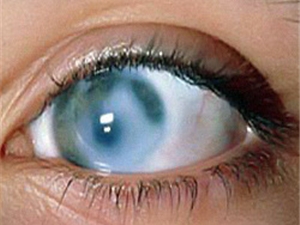
Thus, the most important feature of diabetes is increasing blood sugar levels known also as hyperglycemia. The content of sugar in the blood of healthy human fasting 3.33 – 5.55 mg / dL.

Also, it is well known that a leading role in carbohydrate metabolism plays a pancreas performs two main functions:

  • exocrine which is production of digestive enzymes and which fall on the pancreatic duct into the duodenum and are involved in digestion: break down proteins, fats and carbohydrates to less complex compounds, and then be absorbed into the bloodstream;
  • internal secretion i.e. hormone in the so called “islets of Langerhans”;

These places in the body are unevenly distributed in the tissue of the pancreas and they distinguish four main types of cells known as Beta cells which produce insulin, Alpha calls which are in charge of glucagon, Delta cells for somatostatin and P cells for pancreatic.

Due to the total number of cells and their parameters for “health”, “full” and “quality” we can work out their insulin dependent cherished numbers of health from 3.33 to 5.55 mmol / l glucose in the blood.
Insulin is seen as the “key” for the penetration of glucose into the cells of the liver, brain, muscles i.e. the organs which are not able to function without glucose properly.
Blood glucose level is maintained in the main two hormones:

  • insulin which is lowering it;
  • glucagon which is increasing it.

If grossly interfere in nature – it severely punishes us diseases. The main factors contributing to, the development of diabetes are:

    1) Refined foods of modern man, is deprived of crude fiber, forced to work the intestine, impeding absorbability of fats, cholesterol, lipids, and consequently prevents the development of atherosclerosis, unloads insulin y apparatus;
    2) humanity has less to move and increase the mass of his body for tens of kilograms;
    3) humanity lives in conditions of constant stress;
    4) simply overeating;
    5) breathing fire and eating masses of chemicals;
    6) adverse effects of some drugs on carbohydrate metabolism;
    7) cells of the pancreas are sensitive to different viruses.

The twentieth century gave people insulin and hypoglycemic agents which can not only extended but really has saved millions of lives and made it possible for people affected by diabetes but just to live, work actively, make love and have children.

Establish a genetic predisposition to diabetes

The frequency of diabetes mellitus type I in the 0,2 – 0, 3% of the population. In families where one child is suffering from diabetes type I, his brother or sister of risk to get diabetes about 5%. In families where the father has Type I diabetes risk of the disease, 5 – 10%. If the mother is ill, the risk of disease in children 2.5 – 5%. The risk of developing type I diabetes mellitus is higher in those families in which there is diabetes, a third child’s risk to get 10%. In the case of type I diabetes mellitus in relatives of 2 nd degree relatives (uncle, aunt, grandchild) the risk of developing diabetes mellitus 1 – 2%.

The frequency of diabetes mellitus type II is higher than type I diabetes. Among the relatives of the patient with diabetes mellitus type II diabetes risk 25 – 30%, and if both parents have type II diabetes, the risk of developing diabetes in their children after the age of 40 years – 65 – 75%.

Folk methods for treatment and relief

1. Salad of beets and carrots with oranges

Requires: 4 beets 1 carrot 4 cloves of garlic, 0.3 liters of water, 1 orange, 1 teaspoon lemon juice, 4 tablespoons sour cream, 2 tablespoons chopped dill.

Preparation. Take raw beets which are cleaned and rubbed on a coarse grater, add lemon juice. Carrot shred straw to be mixed with beets and pour hot water bringing to a boil at the end. Then the stuff has to be cool. Serve with oranges, sour cream, minced garlic, dill.

2. Cowberry juice

Washed berries are to be poured in boiled water (2 cups of water per 700 g of berries) forl 10 minutes to be rub the berries with the pestle and sieve. Juice has to be filled with Xylitol, Sorbitol or saharitom. Is chilled on 1/2–1 glasses a day.

3.Drink “Autumn”

In brine sauerkraut pour tomato juice, squeeze the juice of half a lemon there, add removed from 1 / 4 lemon zest crushed, cooled boiled water, salt to taste. Stir well. Serve chilled.

For 6 servings: 1 / 2 liters of brine, 1 / 2 liters of juice, 1 / 2 lemon, 1 / 2 cup water.

4.Tomato drink with cheese

You need for 1 serving the following: 2 tablespoons of tomato juice, 50 grams grated cheese, 1 / 2 cup of cold milk.

Preparation. In blender pour grated cheese, pour the tomato juice and cold milk and beat for 1 minute. Serve well chilled.

5. Carrot Cocktail

Required for 4 servings: 2 cups of carrot juice, 2 oranges, 1 lemon, 2 teaspoons honey, 1 cup yogurt or sour milk, 4 – 8 cubes of edible ice.

Preparation. In a mixer whip the good carrot, orange and lemon juices, yogurt or fermented baked milk, honey.

Diabetes diet

Posted in Hormones and Metabolism | No Comments »

How to cure glaucoma

October 8th, 2010 by admin

Glaucoma is a formidable and a very serious illness which is equally common among people of different countries no mater they are the inhabitants of the north or south, or from cities and villages, or mental workers towards physical labor ones. The name “glaucoma” comes from the Greek origin. It arose because sometimes the pupil area glaucomatous eyes have a yellowish-green color (In greek “glaukos” means “light green”). This thing happens during an acute attack of glaucoma. Currently, glaucoma is perceived as the main cause of incurable blindness and profound disability. You have to know the fourth part of the blinded from various diseases are patients who have lost vision from glaucoma.

Glaucoma cure

The cardinal sign of glaucoma is violation of the regulation of intraocular pressure. Increased intraocular pressure for the second time leads to the development of all the other symptoms characteristic of the glaucomatous process.

The boundaries of normal intraocular pressure normally ranges from 18 to 27 mm Hg. The so called “top ophthalmotonus” for the most of people is in the early morning and later during the day the intraocular pressure is reduced by 2 – 5 mm Hg.

Glaucoma treatment

The most common treatment of congenital glaucoma is the surgical one. In fact, before that you are supposed to take the drug treatment with 1 – 2% solution of pilocarpine and 2 – 3% solution of atseklidiya or 0.25% solution optimola.

The so called youth glaucoma means that developed glaucoma at a young age. There is a hereditary transmission and usually the sick persons are the ones who are under 30 years. The clinical picture is diverse i.e. in some patients with marked changes in the iris, the other symptoms progressed slowly, the cornea of normal size, anterior chamber depth.

Basic principles of medical treatment of primary glaucoma

  • local application of pilocarpine, optimola, klodilina,
  • in the absence of compensation glaucomatous process for the deterioration of visual function the surgery is needed.

Currently, pharmacological treatment of glaucoma conducted in three main areas:

  • normalization of intraocular pressure (therapy of local and general);
  • treatment, contributing to improve the blood supply of the inner shells eye and optic nerve;
  • treatment aimed at normalizing the metabolism in tissues of the eye to influence the degenerative processes typical of glaucoma.

Secondary glaucoma

Glaucoma may develop at different times after the removal of cataracts. Sometimes glaucoma may be as a manifestation of primary open-angle glaucoma and not diagnosed before cataract extraction. Treatment is done with 1 – 2% solution of pilocarpine joined with 0,25 – 0, 5% solution of clonidine 2 – 3 times per day and a 50% solution of glycerol or glycerin with ascorbic acid and fruit syrup.

Secondary glaucoma, disturbed circulation in the vessels of the eye, orbit and intraocular hemorrhages occur more frequently in the central vein thrombosis of the retina, with fewer violations of the venous circulation in the orbit (inflammation, swelling, throbbing enzoftalmy, edematous exophthalmos), with obliteration of the veins after diathermy coagulation of the sclera and its resection, with intraocular hemorrhages.

Therapeutic agents can help relieve hemorrhage: local – instillation of 3% solution of potassium iodide; 0,1% aqueous solution of lidasa or ronidazy; intramuscular use of lidasa, vitreous body.

Posted in Eye | No Comments »

How to cure conjunctivitis

October 8th, 2010 by admin

Conjunctivitis disease comprise about one third of all eye diseases. Amongst them the leading place is in favor of inflammatory infectious conjunctivitis.

Acute and chronic conjunctivitis may have contagious nature caused by viruses and bacteria. Well, there may be also conjunctivitis caused by physical and chemical hazards as well as allergic conjunctivitis and conjunctivitis in common diseases.

Infectious conjunctivitis have expressed greater prevalence of contagious especially in kindergartens, nurseries, schools, educational institutions and industrial enterprises. Conjunctivitis are often caused by staphylococcus also.

Acute conjunctivitis starts first in one but soon comes to the other eye. Patients complain of a sense of debris or so called “sand” in the eye followed by pain and redness in the eyes as well as a watery or mucous-purulent discharge from the eye. Waking up in the morning, the patient normally had difficulty opening his / her eyes as the eyelids glued together with dried pus on the eyelashes. This may be preceded by entering the eye dust, cooling or overheating, swimming in non-flowing water and the general weakening of the organism, diseases of the nose, cold in the chest, etc.

Conjunctivitis cure

For treatment of acute conjunctivitis in order to remove purulent discharge it is necessary to conduct by frequent washing of the eyeball. For the successful treatment they use 2% solution of boric acid and solution furacilinum of 1:5000 or 1:5000 of potassium permanganate solution (pale pink in terms of color). When doing washing the eyelids one should be well diluted and use a rubber spray.

Between washing the conjunctivitis cavity should be done in 2 – 3 hours antibacterial drops. As is often acute conjunctivitis caused coccal flora, the most appropriate to apply the sulfonamides and antibiotics: 30% solution of sodium sulfacyl and 1% solution of tetracycline and 1% solution of tetramitsina.

In acute conjunctivitis in any case it is not possible to apply the bandage on the eye patch since under the bandage blinking is not possible which contributing to the evacuation of the conjunctival cavity of purulent discharge.

Overall treatment normally should be done in a long time period and during the treatment some drugs which have the lack of effectiveness must be replaces in 1 – 2 weeks. You are to use 1% of erythromycin ointment and 1 % emulsion of chloromycetin.

Prevention of acute conjunctivitis is to comply with the rules of personal hygiene and do not touch eyes with dirty hands, do not apply common goods. All persons who were in contact with the patient, prophylaxis is recommended for 2 – 3 days instilled into the eyes of a 30% solution of sodium sulfacyl, in other words albutsid. Also, one should change daily towel and linen until the termination of purulent discharge and it is strictly prohibited to use common items of daily use. Infection can occur from an infected person via dirty hands, as well as by airborne droplets.

Most acute conjunctivitis are caused by adenovirus and may occur in the form of fever with the rise of temperature, inflammation of the upper respiratory tract and increasing the submandibular lymph nodes.

Catarrhal conjunctivitis is the most mild manifestation of the disease and lasts an average of 5 – 7 days. Full recovery might be achieved in 15 days.

Pseudomembranous form of decease is rather rarer. The thin transparent covers are usually soft, grayish-white and easy to remove moist cotton swab. However, in some cases form a fairly dense fibrinous covers with difficulty separating from the subject of inflamed mucosa. After removing the exposed surface of the transparent cover can bleed. Follicular form is characterized by rash follicles or vesicles on the conjunctiva of the cartilage and transitional folds. Follicles may be many in quantities and large and sprinkle the entire loosening mucous membrane century. The disease is very similar to the initial stage of trachoma.

Treatment of conjunctivitis

The following remedies are successfully applied:

  • Florenalum (0,1% solution in drops),
  • Oxolinum (0,1 – 0, 2% in the form of burial),
  • Tebropheum (0,1% solution and 0.5% ointment Nye)
  • Gludantanum (0,1% aqueous solution in drops 6 times a day).

Sulfanilamide (30% solution of sodium sulfacyl) and antibiotics do not have a specific effect on the adenovirus, but they are appropriate for the prevention of secondary infection. The best effect is observed with drugs tetracycline and chloromycetin because these antibiotics have strong virus-stopping effect.

The overall resistance to adenovirus infection increases dibasol, salicylates, vitamins and desensitization drugs.

Сonjunctivitis, caused by physical and chemical hazards, is in most cases professional chronic diseases of conjunctiva. The polluted air, dust, smoke and vapors of various chemicals can cause chronic conjunctivitis. The harmful effect of these agents to be observed in various industrial enterprises here are coal, cement, flour milling, sawmilling, many kinds of chemical industry, where workers have to deal with acids, alkalis, pairs of other compounds.

Complaints of patients include redness, feeling debris eye, tearing, burning, heaviness in the eyelids, growing in the evening. In the morning at the inner corner of eye slit is collected and separated in the form of lumps of mucus.

Treatment of chronic conjunctivitis is more often implies the use of drops and cold compresses of binding agents (infusion of tea, 0.25% solution of resorcinol), with exacerbations and the presence of discharge – 30% solution of sodium sulfacyl and antibiotics. Desinfecting ointments are usually applied nights. In case of professional conjunctivitis it is required to to comply with preventive measures to eliminate the impact of harmfull influence.

Posted in Eye | No Comments »

How to cure vitiligo

October 4th, 2010 by admin

Vitiligo or piebald skin is more often acquired by the disappearance of pigment in normal skin, characterized by the appearance on the skin of different size, shape and localization, depigmented, sharply delineated spots of milky-white color.

Jackson vitiligo

The disease was known in ancient Egypt, but until now the reasons for it remain unclear.

The spots usually appear on the intact skin with gradually increasing and merge forming large foci.

Most affected exposed skin places are: face, neck, hands and skin of large folds, genitals and anus.

The hair on the depigmented areas of skin are usually loosing their normal color too. The skin deprived of pigment is very sensitive to the sun light and under the influence of the sun they quickly turn red, but not tanned and discolored spots in particular stand out against tanned skin.

Folk medicine treatment

Folk medicine recommendationss are various but mainly herbal:

    1. Hypericum perforatum herb – 30,0 gr.
    calendula flowers drug – 30,0 gr.
    Chamomile pharmacy – 20,0 gr.
    Grass Sage – 40,0 gr.
    herb Oregano – 20,0 gr.
    grass succession – 20,0 gr.
    nettle leaves – 40,0 gr.

    From the mixture of herbs you have to prepare infusion as follows: 1 – 2 tbsp. collection pour with 1 cup of hot boiled water then cover and heat on a water bath (note! The water in the bank must be boiling) for 15 minutes then cool at room temperature for 45 minutes. The remaining raw overcome. Volume bring boiled water to 200 ml. Take 1 / 2 cup 2 times a day before meal for 1 – 2 months.

    2. The infusion is prepared the same way as in the first recipe, but has a slightly different composition:
    St. John’s herb – 20,0 gr.
    Calendula officinalis (flowers) – 10,0 gr.
    sage (herb) – 20,0 gr.
    origanum (grass) – 10,0 gr.
    Chamomile pharmacy (flowers) – 10,0 gr.
    Plantago major (leaf) – 15,0 gr.
    nettle (leaf) – 15,0 gr.

    Take a glass of 1/3-1/2. It is highly recommended 3 times daily before meals.

Chinese folk medicine recommends for treatment of small Vitiligo which is floating on the surface of the water plant also known as “duckweed the little”.

You are to take 1 tbsp. of it cleanly washed and chopped and insist duckweed for 3 – 4 days in 1 cup of vodka, then strain. Allow to the patient consume 15 – 20 drops from 2 – 3 tbsp. l. water 2 – 3 times a day.

Also washed crushed duckweed mixed with honey in equal quantities are allowed to be teken1 g 2 – 3 times a day.

Posted in Skin | No Comments »

How to cure rickets

October 1st, 2010 by admin

Rickets is a disease that develops due to lack of vitamin D also known as hypovitaminosis i.e. phosphorus disorders and calcium metabolism, leading to functional disturbance of the Central Nervous System (CNS) as well as changes in bones i.e. joint and muscle systems, accompanied by the development of polyvalent hypovitaminosis.

Rickets bones

Inadequate intake of calcium from food or violation of its absorption in the intestine is accompanied by a decrease in calcium levels, and its ionized fraction in serum. Hypocalcemia stimulates production and secretion of parathyroid hormone which leads to reduction of calcium from bones and increase its level in serum as well as increasing its reflection in the kidneys and increase the synthesis of calcium in the kidneys which is known as Triolet. Homeostasis of phosphate is often regulated by the kidneys i.e. they are absorbed in the intestine almost completely, and their excretion in the urine determines their level in the blood. Excessive absorption of phosphate in the intestine leads to a reduction in serum ionized calcium and increased parathyroid hormone secretion, manifested phosphaturia accompanied by a decrease in phosphate content in the serum and the increase in the amount of calcium. Hypophosphataemia blocks the secretion of parathyroid hormone and activates the synthesis of the calcitriol in the kidneys. This connection enhances the absorption of phosphate in the intestine. Understanding the metabolism of vitamin D is necessary for the understanding of rickets.

The skin contains an enzyme that is under the influence of ultraviolet radiation is converted into vitamin D3. Unavailability of skin to ultraviolet radiation due to smog or clothing also leads to rickets.

There are seven natural substances containing vitamin D with activity but in medical practice they use only 2 of them known as vitamin D2 – called “calciferol” and D3 which is called “cholecalciferol”. In the organism the vitamin itself does not function but its active metabolites, in particular calcitriol.

Calcitriol and other vitamin D metabolites penetrate cell membranes and in cells of organs targeting and interact with specific receptors, forming a complex with them, penetrating into the nucleus of cells and initiates protein synthesis in them. Proteins may be specific (proteins, calcium binding – BSK) and nonspecific – collagen, alkaline fosfatoza. Formed in the brush rim mucosal cells of intestine alkaline fosfatoza participates in an active capturing of calcium ions from the intestinal lumen into the cell with which it interacts with the protein binds calcium, facilitating the passage of calcium through the gut wall into the blood.

Under the influence of metabolites of vitamin D in the intestinal-specific form factors which are also necessary stuff for the absorption of phosphate and magnesium ions. The protein binds calcium circulating in the blood, helps transport calcium in the tissue. In the bones under the influence of calcium Triola is synthesized protein binding calcium, alkaline fosfatoz and normal collagen structure. In the diaphysis kaltsiytriol intensifying resorption of bone tissue it is demonstrated in increasing intake of calcium acetate in blood plasma, which in the kidney is filtered into the primary urine, and from it and then reabsorbed (with normal vitamin D in the body), maintaining a healthy level and citrate, calcium and blood . Resorption of bone with normal vitamin A in the body occurs with a low rate in terms of the resumption of bone tissue. In the kidneys under the influence of calcitriol and is synthesized protein binding calcium, alkaline phosphatase, required for the reabsorption in the proximal tubules of kidneys of calcium, sodium, phosphate, amino acids.

Deficiency of mineral salts violates bone mineralization. Their failure in the areas of growth, shown slowdown and lag bone age is called rickets. The lack of mineralization of trabecular bone means leading to a high content nonmineralized osteoid which is called osteomalacia. Rickets is sick only a growing child and the process in this case is localized mainly in the epiphyses and osteomalacia can occur at any age. All patients revealed osteomalacia rickets, but not all patients with osteomalacia rickets develops.

The disease can occur as a background of deficiency of calcium or phosphorus. Since the ions of calcium and phosphorus are the mineral part of bone, the lack of any of them in the extracellular fluid bathing the mineralized surface, leads to rickets.

Periods of disease are: initial, peak, recovery, residual effects.

The initial stage of the disease accounts for up to the third month of life and shows changes in the nervous system: the child becomes irritable, often crying, sweating, there is a disorder of the fecalium and balding. This period lasts 2 – 3 weeks. In the blood decreases the amount of phosphorus with alkaline phosphatase is activated. In the midst of illness the changes in bone occur: strain softening of skull bones, soften the edges, sternum forward. As a result we see the chest being deformed, deformed limb bones, bone tissue becomes brittle and along with rickets osteoporosis. The X-ray shows not just proper bones but the contours of the bones lubricated. Tubular bone-shaped, fringed edge of the metaphysis. When rickets II – III degree enlarged liver and spleen, expressed anemia.

In acute course has been a rapid increase in symptoms of rickets. Acute course in children during the first months of life, especially in preterm good gain in weight.

Pathogenetic mechanism lies in the violation of phosphate reabsorption in the proximal tubules of the kidneys, the transformation of the inactive metabolite of vitamin D in the active form. Left untreated, the overall body length of an adult does not exceed 130 – 160 cm.

Radiographic signs are to enhance and destruction metaphysis, rough structure of trabecular bone. Goblet change occurs in the metaphysis of the proximal and distal tibia, radius and ulna bones.

Laboratory tests: serum calcium levels did not change or slightly decreased (90 – 94 mg per liter), moderately reduced levels of phosphate (15 – 30 mg / l) and increased activity of alkaline phosphatase. Phosphate excretion in the urine is significant.

The earlier a child develops kidney failure, the longer its course and so more likely to develop osteodystrophy. The early symptoms include slow growth as a result of metabolic acidosis, inadequate diet, a violation of mineral metabolism. Growth retardation sometimes occurs without radiological changes of the skeleton. With progression of the disease develop muscle weakness, bone pain, bone deformities, fractures and epiphyseal displacement in the metaphysis. Particularly noticeable in small children varus curvature of the knee joint, protruding frontal bone, pathological changes of teeth.

Unfortunately, currently the most common type of rickets we face is a classic D i.e. deficient rickets. This is due to a lack of vitamin D from food to both the mother and her child, in connection with the socio – political upheaval, amazing our country.

Important!
Ti is of great importance in the treatment of hypervitaminosis. Patients need to consume vitamin E, stabilizing cell membranes and vitamin A which is able to block the entry of calcium in the tissue. Should be the take of Furosemide, which accelerates the excretion of calcium in the urine. At the same time usually drugs are prescribed drugs that increase the alkaline reserves, liquidate acidosis (sodium bicarbonate, etc.) to be combined with Asparcam and a diet with restriction of calcium.

Posted in Bones and Muscles | No Comments »

 
© 2024 Doctor Medic Powered by Wordpress
="clearer">